Αρχειοθήκη ιστολογίου

Κυριακή 15 Απριλίου 2018

Impact of Powered and Tissue-Specific Endoscopic Stapling Technology on Clinical and Economic Outcomes of Video-Assisted Thoracic Surgery Lobectomy Procedures: A Retrospective, Observational Study

Abstract

Introduction

Video-assisted thoracic surgery (VATS) lung resections are complex procedures with a critical role played by endoscopic staplers in the transection of vessels, bronchi, and lung tissue. This retrospective, observational study compared hospital resource use, costs, and complications of VATS lobectomy procedures for whom powered versus manual endoscopic surgical staplers were used.

Methods

Patients ≥ 18 years of age undergoing elective VATS lobectomy during an inpatient admission from January 1, 2012 to September 30, 2016 were identified from the Premier Healthcare Database (first admission = index admission). Use of either powered or manual endoscopic staplers during the index admission was identified from hospital administrative records. Multivariable regression analyses adjusting for patient, hospital, and provider characteristics and hospital-level clustering were carried out to compare the following outcomes between the powered and manual stapler groups: hospital length of stay (LOS), operating room time (ORT), hospital costs, complications (bleeding and/or transfusions, air leak complications, pneumonia, and infection), discharge status, and 30-, 60-, and 90-day all-cause readmissions.

Results

The powered and manual stapler groups comprised 659 patients (mean age 66.1 years; 53.6% female) and 3100 patients (mean age 66.7 years; 54.8% female), respectively. In the multivariable analyses, the powered stapler group had shorter LOS (4.9 vs. 5.9 days, P < 0.001), lower total hospital costs ($23,841 vs. $26,052, P = 0.009), and lower rates of combined hemostasis complications (bleeding and/or transfusions; 8.5% vs. 16.0%, P < 0.001) and transfusions (5.4% vs. 10.9%, P = 0.002), compared with the manual stapler group. Other outcomes did not differ significantly between the study groups. Similar trends were observed in subanalyses comparing devices across predominant manufacturers in each group, and in subanalyses of patients with comorbid chronic obstructive pulmonary disease.

Conclusion

In this analysis of VATS lobectomy procedures, powered staplers were associated with significant benefits with respect to selected types of hospital resource use, costs, and clinical outcomes when compared with manual staplers.

Funding

Johnson & Johnson.


from #ORL-AlexandrosSfakianakis via ola Kala on Inoreader https://ift.tt/2vgnH03
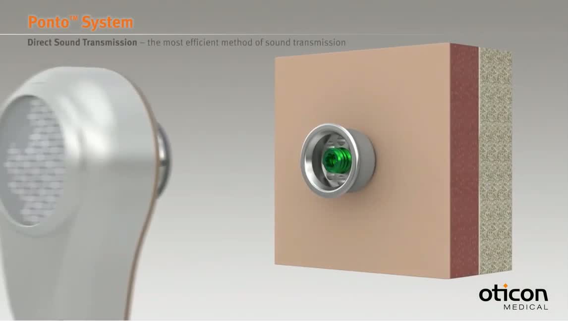
Bone Anchored Hearing Systems - Principles and Candidacy

A review of Bone Anchored Hearing System patient indications, benefits, and candidacy, as well as considerations for selecting and fitting these systems.

from #ORL-AlexandrosSfakianakis via ola Kala on Inoreader https://ift.tt/2qyjvns

CBNAAT: A Boon for Early Diagnosis of Tuberculosis-Head and Neck

Abstract

Tuberculosis of head and neck has been an under diagnosed entity due to large number of smear negative cases, which results in missing out the positive cases, further increasing the burden of TB. The role of cartridge- based nucleic acid amplification test (CBNAAT) with a potential to diagnose TB and rifampicin resistance within 2 h is promising. The study highlights the extended implications of CBNAAT in infectious lesions of head and neck, where the pus or aspirate was subjected to this test, along with other investigations which have been routinely used for detection of extra pulmonary tuberculosis. Twelve patients with infective lesions of head and neck were included in this prospective study, conducted in Department of Otorhinolaryngology, Netaji Subhash Chandra Bose Medical College and hospital, Jabalpur from September 2016 to March 2017. They were investigated for pulmonary and extra pulmonary TB. CBNAAT, microscopy, FNAC and HPR from the site of lesion were done. Nine out of twelve patients were diagnosed positive for Tuberculosis. Microscopy (ZN staining) could detect only two such cases, whereas FNAC showed granulomatous lesion in 3 cases (33.3%). CBNAAT was positive in 77.7% of the total positive cases. Histopathological examination showed 100% results but was feasible only in selected number of cases (4 in this study). CBNAAT provides a promising role in early diagnosis of TB in head and neck. Its high sensitivity and less time taking procedure makes it an excellent tool for timely diagnosis of such cases.


from #ORL-AlexandrosSfakianakis via ola Kala on Inoreader https://ift.tt/2JPRNL6

Thyroid Dysfunction Following Management of Non-thyroid Head and Neck Cancers

Abstract

Head and neck cancers are one of the commonest malignancies in India. Majority of cases of head and neck malignancy undergo chemoradiation with or without surgery. Thyroid bears the brunt in terms of either excision or the gland tends to get irradiated and fibrosed. In either scenario the functionality of gland is lost leading to hypothyroidism and other clinical manifestations. It tends to get subclinical and goes unnoticed. To identify the occurrence of clinical and subclinical hypothyroidism among head and neck cancer patients receiving radiation to the neck and to justify routine use of thyroid function tests during follow up. It was a prospective non randomized control study of 100 patients of head and neck cancer receiving radiotherapy for duration of 1 year. Thyroid stimulating hormone and T3 and T4 estimations were done at baseline and at 3 and 9 months following radiotherapy. Out of 100 patients, 72 (72%) were males and 28 (28%) were females. All the patients received radiation to the neck to a dose of > 30 Gy. 35 patients received concurrent chemotherapy. 11 patients were found to have subclinical hypothyroidism while 32 patients developed significant clinical hypothyroidism (P value of 0.001). Thus a total of 43 patients developed radiation induced hypothyroidism. 20 of the 32 patients who developed clinical hypothyroidism were in the age group of 41–50 years. 11 of 32 patients who developed clinical hypothyroidism received chemoradiation while rest 21 received radiotherapy alone. Mean period for developing radiation induced hypothyroidism was 4.5 months. Hypothyrodism (clinical or subclinical) is an under recognised morbidity of external radiation to the neck which is seen following a minimum dose of 30 Gy to the neck. Recognising hypothyroidism (clinical or subclinical) early and treating it prevents thyroid dysfunction related complications. Hence, thyroid function tests should be made routine during follow up in all patients undergoing radiotherapy.


from #ORL-AlexandrosSfakianakis via ola Kala on Inoreader https://ift.tt/2EMHX8Y

The Reliability of Polyvinylidene Fluoride Sensor for Intra- and Intersession Measurements

Abstract

A new nasal sensor has been designed using Polyvinylidene fluoride (PVDF) film using its piezoelectric property to measure nasal patency. The aim of this study is to determine the intra- and intersession reliability of the new PVDF nasal sensor measurement of unilateral and combined nasal parameters in a group of healthy subjects. Two identical nasal sensors: for right nostril (RN) and left nostril (LN) were designed using piezoelectric natured PVDF films. Twenty subjects were studied. To evaluate the repeatability, total three sets of PVDF sensor measurements were recorded, two sets were taken 5 min apart during same session without repositioning the PVDF nasal sensors and two more sets were taken during 1 h apart successively, by repositioning the PVDF nasal sensor. Intraclass correlation coefficients (ICC) of PVDF sensor measurements for intra- and intersession showed a high and greater repeatability over time for all the combined (mean) and unilateral (RN and LN) values. In both healthy and patients, ICC values for both intra- and intersession measurements were ≥ 0.80 confirming strong reliability and also almost all of the coefficients of variation for the same parameters were low (below 10%). PVDF sensor measurements showed good intra- and intersession repeatability and can be recommended for the objective monitoring of nasal patency during diagnosis and follow-up of conditions.


from #ORL-AlexandrosSfakianakis via ola Kala on Inoreader https://ift.tt/2JNzztz

A Study on Neck Nodes in Oral Cancers, with Special Reference to Skip Metastasis

Abstract

To analyse the distribution of Neck metastases (NM) and to study frequency of skip metastases in oral squamous cell carcinoma (SCC) of oral cavity. From September 2012 to April 2013, 30 previously untreated patients with SCC of oral cavity underwent primary surgical treatment in our institution. From pathological report of Neck dissection specimen prevalence and distribution of NM were ascertained. All patients were classified according to American Joint Committee on Cancer 2005 TNM classification. Overall frequency of NM was 36.7%. Frequency of occult metastases was 33.3%. N+ metastases found in 37% cases. The overall frequency of NM in level IV and V was 9.5%. Isolated level III involvement was found in 3.3%. No isolated level IV and V involvement was found. Skip metastases to level III LN was 6.7%. We did not find any skip metastases to level IV in our study. Neck nodes at greater risk for metastases were level I and II (50 and 28.6%). Level III (11.9%), IV (7.14%), V (2.38%). The risk of skip metastases to level IV was nil in our study.


from #ORL-AlexandrosSfakianakis via ola Kala on Inoreader https://ift.tt/2viqQMz

Effect of photobiomodulation therapy on postoperative pain after endodontic treatment: a randomized, controlled, clinical study

Abstract

Objective

The aim of this prospective, randomized, clinical study was to assess the effect of photobiomodulation therapy (PBM) with low-level laser irradiation (LLLI) on postoperative pain after endodontic treatment.

Materials and methods

Sixty patients, diagnosed with irreversible pulpitis in lower molar teeth, participated in the study. All treatments were performed by a single operator. Participants were randomly divided into two groups: in the experimental group (EG), endodontic treatment was performed with a reciprocating system, immediately followed by PBM with LLLI; and only endodontic treatment was performed in the control group (CG). Postoperative pain was assessed by a second examiner, who was blinded, using two scales: verbal rating scale (VRS) and numerical rating scale (NRS). Assessment was carried out at 6, 12, and 24 h after treatment. Data were analyzed using chi-squared, Fisher's exact, Mann-Whitney tests, ordinal, and non-parametric regression analyses.

Results

For the prevalence of pain, the difference between the groups was significant for the evaluations performed after 6 h (p = 0.04) and 24 h (p = 0.02). The difference after 24 h remained significant after stratification by sex and extrusion of filling material. Increased pain intensity was associated with extrusion of root canal filling material to the periapical region in the two scales used.

Conclusion

The effect of PBM therapy after endodontic treatment showed a significant decreasein prevalence of postoperative pain.

Clinical relevance

The PBM reduces the prevalence of postoperative pain and may benefit patients who need endodontic treatment.


from #ORL-AlexandrosSfakianakis via ola Kala on Inoreader https://ift.tt/2JPg6Zl

Nine prophylactic polishing pastes: impact on discoloration, gloss, and surface properties of a CAD/CAM resin composite

Abstract

Objectives

To investigate discoloration reduction and changes of surface properties of a CAD/CAM resin composite after 14 days´ storage in red wine and polishing with nine different prophylactic polishing pastes (PPPs).

Materials and methods

Rectangular discs (N = 172) were fabricated and polished (P4000) using GC Cerasmart (GC Europe) to investigate different polishing protocols with 1–4 related descending PPPs (22 in total): Cleanic/CLE-Kerr, CleanJoy/CLJ-Voco, Clean Polish/Super Polish/SPO-Kerr, Clinpro Prophy Paste/CPP-3M, Détartrine/DET-Septodont, Nupro/NUP-Dentsply Sirona, Prophy Paste CCS/CCS-Directa, Proxyt/PXT-Ivoclar Vivadent, and Zircate/ZIR Prophy Paste-Dentsply Sirona. Surface properties (roughness values (RV)/Ra, Rz, Rv, surface free energy (SFE), surface gloss (G), and discoloration (ΔE)) were analyzed before and after storage and additional polishing. Data were examined using Kolmogorov-Smirnov test, three-way ANOVA followed by Tukey-B post hoc, Mann-Whitney U, and Kruskal-Wallis H tests (α < 0.05).

Results

Regarding RV, CLE, followed by CCS, and CPP showed the highest values; the lowest presented SPO and DET (p < 0.001). No impact of PPP was observed on ΔE values (p = 0.160). The lowest SFE presented DET, followed by SPO; highest showed CCS followed by NUP and CPP (p < 0.001). Within G, lowest values were observed for CLE and NUP, followed by CCS, ZIP, and CLJ (p < 0.001); the highest presented SPO (p < 0.001). Polishing showed generally a positive impact on SFE values (p < 0.001–p = 0.007), except ZIP (p = 0.322) and CLE (p = 0.083). G increased and RV decreased after polishing (p < 0.001), except SPO, with no significant change for G (p = 0.786).

Conclusions

Polishing with PPPs improves the surface properties and is generally recommended. The choice of PPP has a minor role in removing discolorations. Multi-step systems should be carried out conscientiously.

Clinical relevance

The proper selection of PPP is essential for the clinical outcome of surface properties of prosthetic restorations. Not every polishing paste leads to the same final surface quality.


from #ORL-AlexandrosSfakianakis via ola Kala on Inoreader https://ift.tt/2EPhsj9

Smoking is predictive of poorer distant metastasis-free and progression free-survival in soft tissue sarcoma patients treated with pre-operative radiotherapy or chemoradiotherapy

Abstract

Background

Soft tissue sarcomas (STS) are often treated with pre-operative radiation (RT), with or without chemotherapy, followed by wide local excision. Prognosis for these patients involves an interplay of tumor and patient characteristics. Known prognostic determinants include tumor size, grade, response to therapy, and patient characteristics such as age. While smoking is negatively correlated with outcomes in various malignancies, the impact on STS is unknown. We aimed to assess if smoking impacts overall (OS), distant metastasis-free (DMFS), and progression-free (PFS) survival in patients with STS treated with pre-operative RT.

Methods

Between 2000 and 2015, 166 patients with STS were identified from our prospective database. Patient variables were retrospectively reviewed. Smoking was defined as a ≥ 10 pack year history of current and former smokers. Survival was evaluated using the fisher exact test for univariate (UVA) and logistic regression for multivariate (MVA) analysis.

Results

Fifty-seven (34.3%) patients had smoking histories of ≥ 10 pack years. On UVA, smoking was associated with decreased DMFS (p = 0.0009) and PFS (p = 0.0036), but not OS (p = 0.05). Smoking held significance on MVA for both DMFS and PFS. Current smokers and patients with ≥ 24-month follow-up demonstrated decreased DMFS and PFS on UVA and MVA.

Conclusions

Current smokers and patients with a significant smoking history demonstrated decreased DMFS and PFS in STS patients treated with pre-operative RT. Smoking may cause immunologic compromise and therefore lead to higher rates of progression and distant metastasis.


from #ORL-AlexandrosSfakianakis via ola Kala on Inoreader https://ift.tt/2qAstj7

Role of intralesional bleomycin and intralesional triamcinolone therapy in residual haemangioma following propranolol

Publication date: Available online 14 April 2018
Source:International Journal of Oral and Maxillofacial Surgery
Author(s): V. Pandey, P. Tiwari, S.P. Sharma, R. Kumar, O.P. Singh
With the emergence of propranolol as the first choice of treatment for problematic infantile haemangioma at many centres, the number of patients with a partial or non-response to propranolol has also been growing. This study investigated the role of intralesional bleomycin and triamcinolone in patients with residual disease following propranolol therapy for infantile haemangioma. Sixty-seven patients with residual haemangioma were assigned randomly to receive either intralesional bleomycin (group A, n=36) or intralesional triamcinolone (group B, n=31). The response to treatment and adverse effects were assessed in both groups. All patients received at least four doses and a maximum of six doses of the assigned drug. In group A (mean follow-up 9.38months), 47.2% had an excellent response and 44.4% a good response. In group B (mean follow-up 7.42months), 25.8% had an excellent response and 48.4% a good response. There was no difference in overall response between the groups (P=0.074). Among patients who were initially non-responders to propranolol, bleomycin showed a better response than triamcinolone (P=0.037). This may be due to an overlap in the mechanism of action of propranolol and triamcinolone. Thus, intralesional bleomycin should be preferred in patients with no initial response to propranolol therapy, while bleomycin or triamcinolone can be used in patients with a partial response to propranolol therapy, as they have equal efficacy.


from #ORL-AlexandrosSfakianakis via ola Kala on Inoreader https://ift.tt/2H0ri7M

Otometrics Unveils the Next Generation Bio-logic solutions

OTO_Bio-logic_baby_web.pngOtometrics has introduced the next generation Bio-logic® solutions. Bio-logic has been the trusted brand among professionals for easy-to-use and accurate hearing diagnostics and screening since 1979. Natus Medical Incorporated acquired Bio-logic in 2006 and today Otometrics is reintroducing this legacy brand as part of its global portfolio. "Bio-logic is a well-loved brand with a proud legacy of providing reliable testing," said Mona Dworsack, AuD, Global Director of Product Management, Hearing Assessment and Screening."Our innovation is inspired by the professional's work flow. Our next generation Bio-logic products offer a range of test combinations in a single device – so it's easy to adapt to changing patient needs." 

Bio-logic diagnostic solutions are especially well suited for audiologists who need space-saving devices that are efficient and flexible. Powered by advanced modular technology, the next generation Bio-logic allows screeners to choose a test combination to suit the needs of their clinic​. Four new Bio-logic products will be on display at AAA 2018 booth #621. The products are available for sale in select countries at the conclusion of the conference. Existing Bio-logic users and Otometrics customers are encouraged to contact their local Otometrics/Audiology Systems representatives to start customizing their next generation Bio-logic solution. 
Published: 4/13/2018 5:50:00 PM


from #ORL-AlexandrosSfakianakis via ola Kala on Inoreader https://ift.tt/2H0vNPM

Eosera to Expand Cerumen Removal Product Line

​Eosera (https://earcaremd.com/), the maker of Earwax MD, is releasing new cerumen-targeted products, and will be offering a sneak peek at all the new products at AudiologyNow! 2018 in Nashville, TN. Among Eosera's new products to be unveiled is WaxBlaster MD, a rinsing device to be used after cleaning the ear canal with Earwax MD. Elyse Dickerson, co-founder and CEO of Eosera, said the market has responded well to Earwax MD since last year when it was first introduced. "In retailers carrying the product, dollar sales are up over 18 percent in the ear care category. This growth is primarily driven by the introduction of Earwax MD," Dickerson said. Eosera will sell the products directly and via various resellers for sale in-clinic. At AudiologyNow! 2018, attendees can find out more about Earwax MD and Eosera's new products, including WaxBlaster MD, at Booth 434​. 

Published: 4/13/2018 5:47:00 PM


from #ORL-AlexandrosSfakianakis via ola Kala on Inoreader https://ift.tt/2qAAVPD

Promising drug may deter hearing loss, study finds

A new drug has the potential to revolutionize the prevention and treatment of hearing loss resulting from noise, drug toxicity, and possibly aging, researchers at St. Jude Children's Research Hospital claims. The researchers discovered inhibitors of enzyme Cyclin-dependent kinase 2 (CDK2) that may protect people from developing hearing loss, including hearing impairment in the elderly population.

hear.JPGThe study's lead author Jian Zuo, PhD told The Hearing Journal, "We have reported a genomic study that identified a predisposition to cisplatin-induced hearing loss (CIHL) among pediatric brain tumor patients (Xu et al., Nature Genetics 2015). While predicting which patients will develop CIHL is important, it is more imperative to develop drugs to prevent CIHL. There are no FDA-approved drugs for hearing loss. It is these thoughts that inspired us to screen for drugs against CIHL."

The researchers developed an approach to mimic mammalian cochlear cell death caused by antibiotics, noise, aging, and cisplatin to discover otoprotectants. Cisplatin is a chemotherapy agent, a treatment for an array of cancers, reported to cause permanent hearing loss in patients.

Examining a bioactive library of more than 4,000 unique compounds of an immortalized cell from a cochlear cell line of a neonatal mouse, study authors found ten compounds with protective effects against cisplatin ototoxicity. Among the top-hit compounds was kenpaullone, an inhibitor of CDK2 and other kinases, which are found in zebrafish, adult mice, and rats. Kenpaullone was found to deter cisplatin- and noise-induced damage.

"Our compounds are protective [not only] against CIHL but also noise-induced hearing loss (NIHL). It is also likely that it protects against age-related hearing loss (ARHL)," Zuo explained. "Both NIHL and ARHL affect a large fraction of the society. Based on our results in an identical assay, our compound exhibits better protection against cisplatin-induced cell loss than four benchmark compounds currently in clinical trials. Therefore we believe that our compound will eventually fare well in future clinical trials."

When asked if the CDK2 inhibitors can help patients with inborn hearing loss, Zuo explained, "Our compounds will protect HL but have not been shown to restore hearing among patients with congenital deafness."

The researchers have filed a patent on the methods and the compositions of the CDK2 inhibitors that are discovered to prevent cisplatin- and noise-induced hearing loss.

Published: 4/10/2018 1:27:00 PM


from #ORL-AlexandrosSfakianakis via ola Kala on Inoreader https://ift.tt/2H0dlXr

Adenoid cystic carcinoma of head and neck: A retrospective clinical analysis of a single institution.

Related Articles

Adenoid cystic carcinoma of head and neck: A retrospective clinical analysis of a single institution.

Auris Nasus Larynx. 2018 Apr 10;:

Authors: Chang CF, Hsieh MY, Chen MK, Chou MC

PMID: 29653784 [PubMed - as supplied by publisher]


from #ENT-PubMed via ola Kala on Inoreader https://ift.tt/2qEemuj

Real-world use, safety, and survival of ipilimumab in metastatic cutaneous melanoma in The Netherlands

Phase III trials with ipilimumab showed an improved survival in patients with metastatic melanoma. We evaluated the use and safety of ipilimumab, and the survival of all patients with metastatic cutaneous melanoma (N=807) receiving ipilimumab in real-world clinical practice in The Netherlands using data from the Dutch Melanoma Treatment Registry. Patients who were registered between July 2012 and July 2015 were included and analyzed according to their treatment status: treatment-naive (N=344) versus previously-treated (N=463). Overall, 70% of treatment-naive patients and 62% of previously-treated patients received all four planned doses of ipilimumab. Grade 3 and 4 immune-related adverse events occurred in 29% of treatment-naive patients and 21% of previously-treated patients. No treatment-related deaths occurred. Median time to first event was 5.4 months [95% confidence interval (CI): 4.7–6.5 months] in treatment-naive patients and 4.4 months (95% CI: 4.0–4.7 months) in previously-treated patients. Median overall survival was 14.3 months (95% CI: 11.6–16.7 months) in treatment-naive patients and 8.7 months (95% CI: 7.6–9.6 months) in previously-treated patients. In both patient groups, an elevated lactate dehydrogenase level (hazard ratio: 2.25 and 1.70 in treatment-naive and previously-treated patients, respectively) and American Joint Committee on Cancer M1c-stage disease (hazard ratio: 1.81 and 1.83, respectively) were negatively associated with overall survival. These real-world outcomes of ipilimumab slightly differed from outcomes in phase III trials. Although phase III trials are crucial for establishing efficacy, real-world data are of great added value enhancing the generalizability of outcomes of ipilimumab in clinical practice. *Anouk Jochems and Brenda Leeneman contributed equally to the writing of this article. Correspondence to Anouk Jochems, MD, MSc, Department of Medical Oncology, Leiden University Medical Center, Albinusdreef 2, PO Box 9600, 2300 RC Leiden, The Netherlands Tel: +31 71 5263486; fax: +31 71 5264036; e-mail: a.jochems@lumc.nl Received October 5, 2017 Accepted March 15, 2018 Copyright © 2018 Wolters Kluwer Health, Inc. All rights reserved.

from #ORL-AlexandrosSfakianakis via ola Kala on Inoreader https://ift.tt/2qyUYPj

Congenital Accessory Palpebral Fissure and Eyelid in a Newborn: A Case Report

The occurrence of an accessory palpebral fissure and eyelid is an extremely rare phenomenon. An isolated accessory palpebral fissure and eyelid have been reported only twice in the literature, and in one case as an extension of Delleman syndrome, or oculocerebrocutaneous syndrome. The authors report a case of a full-term newborn who presented with an accessory palpebral fissure and eyelid associated with microcornea, skin polyps and tags, cutis dysplasia, and hypoplasia of the corpus callosum with an otherwise normal systemic workup and negative genetic screening. Detailed surgical management and histopathological analysis of the accessory findings are also described. Accepted for publication February 21, 2018. The authors have no financial or conflicts of interest to disclose Address correspondence and reprint requests to Robert A. Goldberg, M.D., Stein Eye Institute, 300 Stein Plaza, 1st Floor, Los Angeles, CA 90095. E-mail: Goldberg@jsei.ucla.edu © 2018 by The American Society of Ophthalmic Plastic and Reconstructive Surgery, Inc., All rights reserved.

from #ORL-AlexandrosSfakianakis via ola Kala on Inoreader https://ift.tt/2vgkehS

Similarity in Bilateral Isolated Internal Orbital Fractures

Purpose: In evaluating patients sustaining bilateral isolated internal orbital fractures, the authors have observed both similar fracture locations and also similar expansion of orbital volumes. In this study, we aim to investigate if there is a propensity for the 2 orbits to fracture in symmetrically similar patterns when sustaining similar trauma. Methods: A retrospective chart review was performed studying all cases at our institution of bilateral isolated internal orbital fractures involving the medial wall and/or the floor at the time of presentation. The similarity of the bilateral fracture locations was evaluated using the Fisher's exact test. The bilateral expanded orbital volumes were analyzed using the Wilcoxon signed-rank test to assess for orbital volume similarity. Results: Twenty-four patients with bilateral internal orbital fractures were analyzed for fracture location similarity. Seventeen patients (70.8%) had 100% concordance in the orbital subregion fractured, and the association between the right and the left orbital fracture subregion locations was statistically significant (P

from #ORL-AlexandrosSfakianakis via ola Kala on Inoreader https://ift.tt/2JNNKPa

Frontalis Muscle Flap Versus Maximal Anterior Levator Resection as First Option for Patients With Severe Congenital Ptosis

Purpose: To compare 2 surgical techniques (frontalis flap versus maximal anterior levator resection) as first surgical options for the treatment of congenital ptosis with poor levator function in patients younger than 2 years of age with a follow up of 10 years. Methods: A retrospective study of 58 patients (71 eyelids) with severe ptosis and poor levator function who underwent frontalis muscle flap (FMF = 47) or maximal anterior levator resection (ALR = 24) for correction of their ptosis. Eyelid measurements were taken at baseline, 1, 5, and 10 years after surgery. The presence of complications, need for reoperations, and palpebral contour were evaluated. Results: Most patients in both groups required only one surgical procedure with a stable average margin-reflex distance 1 over the 10-year follow-up period in both groups, with no statistically significant difference between the 2 techniques in achieving an adequate palpebral height after one single procedure. Eleven eyelids treated with FMF (23%) and 12 treated with ALR (50%) needed a reoperation, with a statistically significant difference between the 2 techniques. Five ALR patients (21%) and 6 FMF patients (13%) had alterations of eyelid contour. Pop-eyelid and eyelash ptosis were observed in 8% of patients operated with FMF. Conclusion: Good functional and aesthetic results were obtained with both surgical techniques. FMF required fewer reoperations compared with maximal ALR, offering a better long-term result without residual ptosis. Accepted for publication February 4, 2018. Presented at the XXVI SECPOO meeting (Bilbao, 2016), 35º ESOPRS meeting (Athens, 2016), Barcelona Oculoplastics (Barcelona, 2017), BOPSS (London, 2017). The authors have no financial or conflicts of interest to disclose. Address correspondence and reprint requests to LuzMaria Vasquez, M.D., Josep María Lladó 3, Salida 7 Ronda de Dalt. 08035, Barcelona, Spain. E-mail: vasquez@imo.es © 2018 by The American Society of Ophthalmic Plastic and Reconstructive Surgery, Inc., All rights reserved.

from #ORL-AlexandrosSfakianakis via ola Kala on Inoreader https://ift.tt/2ENpM2I

Orbital Inflammatory Syndrome Secondary to Flea Bite

A 34-year-old previously healthy Hispanic male presented to the emergency room complaining of progressive left upper eyelid swelling and pain for more than 2 weeks. He was previously diagnosed and treated for a "pink eye" but failed to improve. He reported a previous "bug bite" around the left lateral canthus a few weeks prior to admission. Computer tomography orbit with contrast showed left exophthalmos, an enhancing left lacrimal gland and orbital inflammatory signs suggestive of possible intraorbital abscess. Intravenous antibiotics did not improve his symptoms. Surgical debridement showed no abscess but inflamed soft tissues and lacrimal gland. Intravenous steroids failed to improve his symptoms. On postoperative day 3, the patient reported that an insect had "jumped" out from his left orbit. Identification of the specimen proved to be a mature flea. Biopsy of the lacrimal gland showed degranulation of eosinophils and foreign body material consistent with probable insect leg parts. Accepted for publication February 19, 2018. Disclosure: Collection and evaluation of protected patient health information was HIPAA-compliant. Address correspondence and reprint requests to Amina I. Malik, M.D., Blanton Eye Institute, Houston Methodist Hospital, 6560 Fannin Street, Suite 450, Houston, TX 77030. E-mail: aimalik@houstonmethodist.org © 2018 by The American Society of Ophthalmic Plastic and Reconstructive Surgery, Inc., All rights reserved.

from #ORL-AlexandrosSfakianakis via ola Kala on Inoreader https://ift.tt/2JKS69W

Repeated Audiometry After Bacterial Meningitis: Consequences for Future Management

Objective: Sensorineural hearing loss is a common sequela of bacterial meningitis. The objective of this study is to delineate the incidence and course of hearing loss after bacterial meningitis. Study Design: Retrospective cohort study. Setting: Tertiary referral center. Patients: Data of 655 patients who suffered from bacterial meningitis between 1985 and 2015 were analyzed. Interventions: None. Main Outcome Measurements: Availability of audiometric data, incidence of hearing loss, and onset and course of hearing loss. Results: In this cohort the incidence of hearing loss (>25 dB) was 28% (95% confidence interval 23–34%). The incidence of profound hearing loss (>80 dB) was 13% (95% confidence interval 10–18%). Normal hearing at the first assessment after treatment for meningitis remained stable over time in all these patients. In 19 of the 28 patients with diagnosed hearing loss, the hearing level remained stable over time. Hearing improved in six patients and deteriorated in two patients. One patient showed a fluctuating unilateral hearing loss. Conclusion: Audiological tests in patients with bacterial meningitis, especially children, should be started as soon as possible after the acute phase is over. As we found no deterioration of initial normal hearing after bacterial meningitis, repeated audiometry seems indicated only for those with diagnosed hearing loss at first assessment. This is an open access article distributed under the terms of the Creative Commons Attribution-Non Commercial-No Derivatives License 4.0 (CCBY-NC-ND), where it is permissible to download and share the work provided it is properly cited. The work cannot be changed in any way or used commercially without permission from the journal. https://ift.tt/OBJ4xP Address correspondence and reprint requests to Marc P. van der Schroeff, Ph.D., Department of Otorhinolaryngology and Head and Neck Surgery, Erasmus Medical Center, Room SP-1455, P.O. Box 2040, 3000 CA Rotterdam, The Netherlands; E-mail: m.vanderschroeff@erasmusmc.nl This research received no specific grant from any funding agency in the public, commercial, or not-for-profit sectors. The authors disclose no conflicts of interest. Copyright © 2018 by Otology & Neurotology, Inc. Image copyright © 2010 Wolters Kluwer Health/Anatomical Chart Company

from #ORL-AlexandrosSfakianakis via ola Kala on Inoreader https://ift.tt/2GYkQhr

Malar Reconstruction with Anterior Bilobed Cheek Flap

Summary: A 66-year-old man had developed a tumor on his right cheek. A biopsy revealed basal cell carcinoma. The ulcerative cancer and zygomatic bone were resected. We designed an anterior bilobed cheek flap to reconstruct the cheek skin defect. The postoperative clinical course was uneventful. This is an open-access article distributed under the terms of the Creative Commons Attribution-Non Commercial-No Derivatives License 4.0 (CCBY-NC-ND), where it is permissible to download and share the work provided it is properly cited. The work cannot be changed in any way or used commercially without permission from the journal. Published online 13 April 2018. Received for publication November 28, 2017; accepted January 30, 2018. Disclosure: The authors have no financial interest to declare in relation to the content of this article. The Article Processing Charge was paid for by the authors. Koichi Ueda, MD, Department of Plastic and Reconstructive Surgery, Osaka Medical College, Takatsuki, Osaka, 569–8686, Japan, E-mail: pla007@osaka-med.ac.jp Copyright © 2018 The Authors. Published by Wolters Kluwer Health, Inc. on behalf of the American Society of Plastic Surgeons. All rights reserved.

from #ORL-AlexandrosSfakianakis via ola Kala on Inoreader https://ift.tt/2H0EGcd

Does Distal Radio-ulnar Joint Configuration Affect Postoperative Functional Results after Ulnar Shortening Osteotomy?

Background: Reverse oblique distal radio-ulnar joint (DRUJ) configuration is assumed to show inferior postoperative results in ulnar-shortening osteotomy due to osteoarthritis, as the joint force pressure in the DRUJ may be increased. An evaluation and comparison of the postoperative functional results with regard to clinical and radiographic signs of arthritis among different DRUJ configurations was carried out retrospectively. Methods: Sixty-two patients after ulnar shortening osteotomy were included. The minimum follow-up was 5 years. Preoperative x-rays were assessed for the DRUJ configuration according to the Tolat classification, whereas postoperative radiographs were evaluated with regard to signs of osteoarthritis using the Kallgren-Lawrence-Score. Functional results were evaluated using the disabilities of the arm, shoulder and hand (DASH) and Mayo Wrist Score and measuring range of motion and grip strength. Results: Significantly better functional results were found in patients with parallel configuration of the DRUJ (Tolat type 1 configuration) with regard to DASH score, grip strength, and supination compared with nonparallel configurations. In the Tolat type 1, configurated DRUJ mean DASH score was 9 compared with 18 in the Tolat type 2 and 3 groups. Apart from supination, no differences were observed in range of motion among groups. Conclusion: Although long-term postoperative range of motion failed to display statistically significant differences between DRUJ configurations except for supination, better results regarding grip strength and DASH scores were seen in a parallel-aligned DRUJ configuration. Although onset of osteoarthritis does not seem to become apparent within the observation period, nonparallel aligned configuration predisposes to inferior results. This is an open-access article distributed under the terms of the Creative Commons Attribution-Non Commercial-No Derivatives License 4.0 (CCBY-NC-ND), where it is permissible to download and share the work provided it is properly cited. The work cannot be changed in any way or used commercially without permission from the journal. Published online 13 April 2018. Received for publication November 5, 2017; accepted March 1, 2018. Disclosure: The authors have no financial interest to declare in relation to the content of this article. The Article Processing Charge was paid for by the authors. Fabian Gilbert, MD, Department of Trauma, Hand, Plastic and Reconstructive Surgery, Julius-Maximilians-University of Würzburg, Oberdürrbacherstr. 6, D- 97080 Würzburg, Germany, E-mail: gilbert_f@ukw.de Copyright © 2018 The Authors. Published by Wolters Kluwer Health, Inc. on behalf of the American Society of Plastic Surgeons. All rights reserved.

from #ORL-AlexandrosSfakianakis via ola Kala on Inoreader https://ift.tt/2qxN1cj

A Systematic Review of Surgical Management of Melanoma of the External Ear

Background: Historically, external ear melanomas have been treated aggressively, due to early perceptions suggesting they had poor prognosis and aggressive biological behavior. More recent evidence has not supported these notions. Methods: We completed a complete review of the literature involving malignant melanoma of the external ear. We then completed a quantitative analysis on seventy-three cases from 8 reports that contained case-level data, assessing factors that influence recurrence, and assessing characteristics of the melanomas based on histological subtype. Baseline and outcomes data for all 20 studies were then compiled but not statistically evaluated. Results: In our subanalysis, patients who had recurrence were significantly more likely to have had wedge resection versus wide-local excision, and those with no recurrence were more likely to have undergone wide local excision. Nodular tumors had significantly greater thickness. Overall, conservative excisions provided excellent outcomes. Conclusions: Conservative treatment for external ear melanoma produces satisfactory outcomes. There is no evidence to support the use of radical amputation and little evidence to support the removal of cartilage or perichondrium. Sentinel lymph node biopsy is warranted only with positive nodes. There is no role for elective neck dissection. The roles for chemo/radiation therapy are unclear and guidelines for other cutaneous melanomas should be followed. This is an open-access article distributed under the terms of the Creative Commons Attribution-Non Commercial-No Derivatives License 4.0 (CCBY-NC-ND), where it is permissible to download and share the work provided it is properly cited. The work cannot be changed in any way or used commercially without permission from the journal. Published online 13 April 2018. Received for publication February 1, 2018; accepted February 20, 2018. Disclosure: The authors have no financial interest to declare in relation to the content of this article. The Article Processing Charge was paid for by the authors. Michael W. Neumeister, MD, Department of Surgery, Institute for Plastic Surgery, Southern Illinois University School of Medicine, 747 North Rutledge - 3rd Floor, P.O. Box 19653 Springfield, IL 62794–9653, E-mail: mneumeister@siumed.edu Copyright © 2018 The Authors. Published by Wolters Kluwer Health, Inc. on behalf of the American Society of Plastic Surgeons. All rights reserved.

from #ORL-AlexandrosSfakianakis via ola Kala on Inoreader https://ift.tt/2GZvZ1G

The Excised Super-thin Skin as a Flap Sizer for Finger and Hand Free-Mini-Flap Reconstruction

No abstract available

from #ORL-AlexandrosSfakianakis via ola Kala on Inoreader https://ift.tt/2qzGwW7

Shoulder Morbidity in Patients after Head and Neck Reconstruction with the Pedicled Supraclavicular Island Flap

Background: The pedicled supraclavicular artery island flap (SCAIF) for reconstruction of the head and neck has been shown to be a pliable alternative to established pedicled flaps, such as the pectoralis major myocutaneous flap. Because there are limited published data regarding shoulder morbidity after SCAIF procedure, we aimed to investigate it with 2 established questionnaires for the upper extremity [Constant-Murley score and Disabilities of the Arm, Shoulder and Hand Outcome Measure (DASH) score]. Methods: The authors designed and implemented a retrospective cohort study of patients who received a defect reconstruction by SCAIF. Analyzed parameters were demographics, comorbidities, donor-site morbidity and shoulder morbidity in terms of range of motion, pain, strength, and daily activities evaluated and compared between the donor site and contralateral arm. Results: Of the 61 consecutively performed head and neck reconstructions with SCAIF, 20 met inclusion criteria (curative intended treatment, head and neck squamous cell cancer, follow-up time more than 4 months). Mean follow-up was 17.3 months (±10.4 months) ranging from 4–35 months. Donor-site complication rate was low with 5% major (surgical revision) and 30% minor complications (conservative management). Overall Constant-Murley-Score (P = 0.334), pain (P = 0.150), overall range of motion (P = 0.861), and strength of the extremity (P = 0.638) of the shoulder receiving a SCAIF showed no significant differences to the contralateral extremity. Mean of Disabilities of the Arm, Shoulder and Hand Outcome Measure score was 32.5 (±28.6). Conclusion: The results of the present study suggest very low shoulder morbidity in patients after SCAIF procedure with no significant functional impairment of the donor shoulder compared with the contralateral side. This is an open-access article distributed under the terms of the Creative Commons Attribution-Non Commercial-No Derivatives License 4.0 (CCBY-NC-ND), where it is permissible to download and share the work provided it is properly cited. The work cannot be changed in any way or used commercially without permission from the journal. Published online 12 April 2018. Received for publication July 30, 2017; accepted December 19,2017. Disclosure: The authors have no financial interest to declare in relation to the content of this article. The Article Processing Charge was paid for by the authors. Supplemental digital content is available for this article. Clickable URL citations appear in the text. Drs. Spiegel and Pilavakis contributed equally to this work. Jennifer Spiegel, MD, Department of Otorhinolaryngology, Head and Neck Surgery, Marchioninistraße 15, 81377 Munich, Germany, E-mail: Jennifer.Spiegel@med.uni-muenchen.de Copyright © 2018 The Authors. Published by Wolters Kluwer Health, Inc. on behalf of the American Society of Plastic Surgeons. All rights reserved.

from #ORL-AlexandrosSfakianakis via ola Kala on Inoreader https://ift.tt/2GYsyYV

Primary Repair of a Complex Panfacial Fracture by Dog Bite

Summary: Facial fractures due to dog attacks have an unknown incidence rate. To date, only 41 cases of canine bite trauma in a pediatric patient, associated with facial fracture, have been reported in the literature. As major species of involving dogs are the American pitbull terrier and rottweiler. Due to the intense kinematics of this trauma, the treatment becomes complex. Thus, attention to the primary repair of such complex lesions ensures satisfactory results, which is the focus of this discussion. The purpose of this review was to analyze how different ways to approach this type of trauma in children for clarification or correct management. In addition, we address the treatment plan of a complex case of panfacial fracture by a canine bite in a 4-year-old patient. According to a review addressed, the main involved are orbit, nasal, and zygomatic. Antibiotic therapy is indicated for infected bite wounds and wounded considerations at risk of infection, with high complexity and when involving important structures such as bones, vessels, and joints. The state of tetanus immunization and the risk of rabies infection should be routinely addressed in the management of the bite wound. This is an open-access article distributed under the terms of the Creative Commons Attribution-Non Commercial-No Derivatives License 4.0 (CCBY-NC-ND), where it is permissible to download and share the work provided it is properly cited. The work cannot be changed in any way or used commercially without permission from the journal. Published online 12 April 2018. Received for publication August 15, 2017; accepted January 23, 2018. Disclosure: The authors have no financial interest to declare in relation to the content of this article. Ricardo Augusto Conci, PhD Oral and Maxillofacial Surgery, Rua Universitária, 1619, Jardim Universitário 85819-110, Cascavel, PR, Brazil, E-mail: ricardo_conci@hotmail.com Copyright © 2018 The Authors. Published by Wolters Kluwer Health, Inc. on behalf of the American Society of Plastic Surgeons. All rights reserved.

from #ORL-AlexandrosSfakianakis via ola Kala on Inoreader https://ift.tt/2qA3Ov7

Statistical Basic Steps to Be Considered on Planning a Research

No abstract available

from #ORL-AlexandrosSfakianakis via ola Kala on Inoreader https://ift.tt/2GYsubD

Association of Academic Physiatrists Federal Funding of Disability and Rehabilitation Research Position Statement

The Association of Academic Physiatrists (AAP) seeks to advocate for policies that are supportive of academic rehabilitation, with elected officials, agency leaders, and other policy makers. Accordingly, the AAP's Public Policy Committee identifies policy issues of importance to the organizational mission, conducts background research on those issues, and develops position statements that articulate the organization's position. These position statements require approval by the Board of Trustees and are used to support advocacy efforts by AAP members. Federal funding for disability and rehabilitation research is an important issue for the AAP and its members. This position statement addresses the need for greater federal funding in this area, better coordination of the agencies providing funding, and an appropriate balance of merit-based funding with funding targeted to building capacity in critical rehabilitation-relevant methodology and content areas. This position paper was reviewed by the Association of Academic Physiatrists Public Policy Committee and the Board of Trustees and approved as a position paper of the Association. John Whyte, MD, PhD, Moss Rehabilitation Research Institute, 50 Township Line Road, Elkins Park, PA 19027, email: jwhyte@einstein.edu Copyright © 2018 Wolters Kluwer Health, Inc. All rights reserved.

from #ORL-AlexandrosSfakianakis via ola Kala on Inoreader https://ift.tt/2H3twTR

A Novel Technique of Ultrasound-Guided Selective Mandibular Nerve Block With a Lateral Pterygoid Plate Approach: A Cadaveric Study

Background and Objectives We aimed to describe a novel technique of ultrasound-guided selective mandibular nerve block with a lateral pterygoid plate (LPP) approach and to assess its feasibility and accuracy in a soft cadaver model. Methods Ten soft cadavers were studied. A curved array ultrasound transducer was applied over 1 side of the face of the cadaver, in an open-mouth position. The transducer was placed transversely below the zygomatic arch and tilted in the caudal-to-cranial direction to identify the boundary of the LPP. The needle was inserted in-plane, in an anterior-to-posterior direction, into the posterior border of the uppermost part of the LPP, and 3 mL of methylene blue was injected. Results Mandibular nerve block was successfully performed in all 10 cadavers using an LPP approach under ultrasound guidance. The mandibular nerve and its branches were seen to be stained with methylene blue in all cadaveric specimens. No accidental injection into the facial nerve or maxillary artery was observed. Conclusions This cadaveric study suggests that this novel technique, using an LPP approach under ultrasound guidance, is helpful for selective mandibular nerve block, with high accuracy and feasibility. Further studies are required to establish its safety and efficacy for clinical application. Clinical Trial Registration This study was registered at the Thai Clinical Trials Registry (ClinicalTrials.in.th), identifier TCTR20160601004. Accepted for publication November 20, 2017. Address correspondence to: Wirinaree Kampitak, MD, Department of Anesthesiology, King Chulalongkorn Memorial Hospital, The Thai Red Cross Society and Faculty of Medicine, Chulalongkorn University, 1873, Rama 4 Road, Pathumwan, Bangkok, 10330, Thailand (e-mail: nutong127@yahoo.com). No external funding was received. The authors declare no conflict of interest. Copyright © 2018 by American Society of Regional Anesthesia and Pain Medicine.

from #ORL-AlexandrosSfakianakis via ola Kala on Inoreader https://ift.tt/2HF7DHK

The Impact of Spinal Needle Selection on Postdural Puncture Headache: A Meta-Analysis and Metaregression of Randomized Studies

Background and Objectives Potentially broadened indications for spinal anesthesia require increased understanding of the risk factors and prevention measures associated with postdural puncture headache (PDPH). This review is designed to examine the association between spinal needle characteristics and incidence of PDPH. Methods Meta-analysis and metaregression was performed on randomized controlled trials to determine the effect of needle design and gauge on the incidence of PDPH after controlling for patient confounders such as age, sex, and year of publication. Results Fifty-seven randomized controlled trials (n = 16416) were included in our analysis, of which 32 compared pencil-point design with cutting-needle design and 25 compared individual gauges of similar design. Pencil-point design was associated with a statistically significant reduction in incidence of PDPH (risk ratio, 0.41; 95% confidence interval, 0.31–0.54; P

from #ORL-AlexandrosSfakianakis via ola Kala on Inoreader https://ift.tt/2GZpdVG

Selective Suprascapular and Axillary Nerve Block Versus Interscalene Plexus Block for Pain Control After Arthroscopic Shoulder Surgery: A Noninferiority Randomized Parallel-Controlled Clinical Trial

Background and Objectives This randomized trial aimed to assess if a combined suprascapular-axillary nerve block (SSB) is noninferior (margin = 1.3 on a 0- to 10-point scale) to interscalene block (ISB) in treating pain after arthroscopic shoulder surgery. Secondary end points included opioid consumption, dyspnea, discomfort associated with muscle weakness, and patient satisfaction. Methods One hundred patients undergoing arthroscopic shoulder surgery were randomized to receive ultrasound-guided ISB (n = 50) or SSB (n = 50). Pain intensity at rest, dyspnea, and discomfort were recorded upon arrival in the recovery room, discharge to the ward, and at 4, 8, and 24 hours after surgery. Piritramide consumption was recorded for the first 24 hours. Patient satisfaction was assessed on the second postoperative day. Results During the first 4 hours after surgery, the difference in mean pain score between SSB and ISB was higher than 2.5 (±0.8). The difference gradually decreased to 1.1 (±1.0) at 8 hours before resulting in noninferiority during the night and at 24 hours. Piritramide consumption was significantly higher in the SSB group in the first 8 hours. The incidence of dyspnea and discomfort was higher after ISB. Treatment satisfaction was similar in both groups. Conclusions Suprascapular-axillary nerve block is inferior to ISB in terms of analgesia and opioid requirement in the immediate period after arthroscopic shoulder surgery but is associated with a lower incidence of dyspnea and discomfort. The difference in pain and opioid consumption gradually decreases as the blocks wear off in order to reach similar pain scores during the first postoperative night and at 24 hours. Clinical Trial Registration This study was registered at ClinicalTrials.gov, identifier NCT02415088. Accepted for publication December 30, 2017. Address correspondence to: Björn Stessel, MD, PhD, Department of Anesthesiology and Pain Treatment, Jessa Hospital, Virga-Jesse Campus, Stadsomvaart 11, 3500 Hasselt, Belgium (e-mail: bjornstessel@hotmail.com). This study is part of the Limburg Clinical Research Program (LCRP) UHasselt-ZOL-Jessa, supported by the foundation Limburg Sterk Merk, Province of Limburg, Flemish Government, Hasselt University, Ziekenhuis Oost-Limburg, and Jessa Hospital. The authors declare no conflict of interest. Authors' contributions: A.N. was responsible for the study design, data collection, and writing of the paper. B.S. was responsible for the study design, data interpretation, table creation, and writing of the paper. P.F.W. was responsible for the data interpretation and writing of the paper. C.D. was responsible for the study design and the writing of the paper. W.C. provided statistical expertise and was responsible for the statistical analyses and figure creation. J.-P.O., I.A., L.J., and J.D. were responsible for the writing of the paper. D.S. conceived of the study and was responsible for the study design, the literature search, execution of all regional blocks, and writing of the paper. Copyright © 2018 by American Society of Regional Anesthesia and Pain Medicine.

from #ORL-AlexandrosSfakianakis via ola Kala on Inoreader https://ift.tt/2HBQazX

Clotting-Factor Concentrations 5 Days After Discontinuation of Warfarin

Background The American Society of Regional Anesthesia and Pain Medicine guidelines recommend discontinuation of warfarin and an international normalized ratio (INR) of 1.2 or less before a neuraxial injection. The European and Scandinavian guidelines accept an INR of 1.4 or less. We evaluated INR and levels of clotting factors (CFs) II, VII, IX, and X 5 days after discontinuation of warfarin. Methods Patients who discontinued warfarin for 5 days and had an INR of 1.4 or less had activities of factors II, VII, IX, and X measured. The primary outcome was the frequency of subjects with CF activities of less than 40%. Results Twenty-three patients were studied; 21 (91%) had an INR of 1.2 or less. In these 21 patients, the median (interquartile range) activities of factors II, VII, IX, and X were 66% (52%–80%), 114% (95%–132%), 101% (84%–121%), and 55% (46%–63%), respectively. Ninety-five percent (99% confidence interval, 69%–99%) had CF activities of greater than 40%. The patient who did not CF activities greater than 40% had end-stage renal disease. Two subjects had an INR of greater than 1.2; the activities of factor II, VII, IX, and X were 37% and 46%, 89% and 105%, 66% and 78%, and 20% and 36%, respectively. Neither patient had CF activities of greater than 40%. Conclusions Based on 40% activity of CFs, patients with INRs of 1.2 or less can be considered to have adequate CFs to undergo neuraxial injections. The number of patients with an INR of 1.3 and 1.4 is too small to make conclusions. Accepted for publication January 30, 2018. Address correspondence to: Honorio T. Benzon, MD, Department of Anesthesiology, 251 E. Huron, St, Feinberg Pavilion, 5–704, Chicago, IL 60611 (e-mail: h-benzon@northwestern.edu). L.V. is retired. This work is attributed to the Departments of Anesthesiology and Medicine, Northwestern University Feinberg School of Medicine, Chicago, IL. Funding support for this study was provided by the Department of Anesthesiology, Northwestern University Feinberg School of Medicine (departmental sources only). The study was presented at Anesthesiology 2016, the annual meeting of the American Society of Anesthesiologists, October 22 to 25, 2016, Chicago, IL. The authors declare no conflict of interest. Copyright © 2018 by American Society of Regional Anesthesia and Pain Medicine.

from #ORL-AlexandrosSfakianakis via ola Kala on Inoreader https://ift.tt/2GYV5tC

Repeated Audiometry After Bacterial Meningitis: Consequences for Future Management

Objective: Sensorineural hearing loss is a common sequela of bacterial meningitis. The objective of this study is to delineate the incidence and course of hearing loss after bacterial meningitis. Study Design: Retrospective cohort study. Setting: Tertiary referral center. Patients: Data of 655 patients who suffered from bacterial meningitis between 1985 and 2015 were analyzed. Interventions: None. Main Outcome Measurements: Availability of audiometric data, incidence of hearing loss, and onset and course of hearing loss. Results: In this cohort the incidence of hearing loss (>25 dB) was 28% (95% confidence interval 23–34%). The incidence of profound hearing loss (>80 dB) was 13% (95% confidence interval 10–18%). Normal hearing at the first assessment after treatment for meningitis remained stable over time in all these patients. In 19 of the 28 patients with diagnosed hearing loss, the hearing level remained stable over time. Hearing improved in six patients and deteriorated in two patients. One patient showed a fluctuating unilateral hearing loss. Conclusion: Audiological tests in patients with bacterial meningitis, especially children, should be started as soon as possible after the acute phase is over. As we found no deterioration of initial normal hearing after bacterial meningitis, repeated audiometry seems indicated only for those with diagnosed hearing loss at first assessment. This is an open access article distributed under the terms of the Creative Commons Attribution-Non Commercial-No Derivatives License 4.0 (CCBY-NC-ND), where it is permissible to download and share the work provided it is properly cited. The work cannot be changed in any way or used commercially without permission from the journal. https://ift.tt/OBJ4xP Address correspondence and reprint requests to Marc P. van der Schroeff, Ph.D., Department of Otorhinolaryngology and Head and Neck Surgery, Erasmus Medical Center, Room SP-1455, P.O. Box 2040, 3000 CA Rotterdam, The Netherlands; E-mail: m.vanderschroeff@erasmusmc.nl This research received no specific grant from any funding agency in the public, commercial, or not-for-profit sectors. The authors disclose no conflicts of interest. Copyright © 2018 by Otology & Neurotology, Inc. Image copyright © 2010 Wolters Kluwer Health/Anatomical Chart Company

from #ORL-AlexandrosSfakianakis via ola Kala on Inoreader https://ift.tt/2GYkQhr

Congenital Accessory Palpebral Fissure and Eyelid in a Newborn: A Case Report

The occurrence of an accessory palpebral fissure and eyelid is an extremely rare phenomenon. An isolated accessory palpebral fissure and eyelid have been reported only twice in the literature, and in one case as an extension of Delleman syndrome, or oculocerebrocutaneous syndrome. The authors report a case of a full-term newborn who presented with an accessory palpebral fissure and eyelid associated with microcornea, skin polyps and tags, cutis dysplasia, and hypoplasia of the corpus callosum with an otherwise normal systemic workup and negative genetic screening. Detailed surgical management and histopathological analysis of the accessory findings are also described. Accepted for publication February 21, 2018. The authors have no financial or conflicts of interest to disclose Address correspondence and reprint requests to Robert A. Goldberg, M.D., Stein Eye Institute, 300 Stein Plaza, 1st Floor, Los Angeles, CA 90095. E-mail: Goldberg@jsei.ucla.edu © 2018 by The American Society of Ophthalmic Plastic and Reconstructive Surgery, Inc., All rights reserved.

from #ORL-AlexandrosSfakianakis via ola Kala on Inoreader https://ift.tt/2vgkehS

Similarity in Bilateral Isolated Internal Orbital Fractures

Purpose: In evaluating patients sustaining bilateral isolated internal orbital fractures, the authors have observed both similar fracture locations and also similar expansion of orbital volumes. In this study, we aim to investigate if there is a propensity for the 2 orbits to fracture in symmetrically similar patterns when sustaining similar trauma. Methods: A retrospective chart review was performed studying all cases at our institution of bilateral isolated internal orbital fractures involving the medial wall and/or the floor at the time of presentation. The similarity of the bilateral fracture locations was evaluated using the Fisher's exact test. The bilateral expanded orbital volumes were analyzed using the Wilcoxon signed-rank test to assess for orbital volume similarity. Results: Twenty-four patients with bilateral internal orbital fractures were analyzed for fracture location similarity. Seventeen patients (70.8%) had 100% concordance in the orbital subregion fractured, and the association between the right and the left orbital fracture subregion locations was statistically significant (P

from #ORL-AlexandrosSfakianakis via ola Kala on Inoreader https://ift.tt/2JNNKPa

Frontalis Muscle Flap Versus Maximal Anterior Levator Resection as First Option for Patients With Severe Congenital Ptosis

Purpose: To compare 2 surgical techniques (frontalis flap versus maximal anterior levator resection) as first surgical options for the treatment of congenital ptosis with poor levator function in patients younger than 2 years of age with a follow up of 10 years. Methods: A retrospective study of 58 patients (71 eyelids) with severe ptosis and poor levator function who underwent frontalis muscle flap (FMF = 47) or maximal anterior levator resection (ALR = 24) for correction of their ptosis. Eyelid measurements were taken at baseline, 1, 5, and 10 years after surgery. The presence of complications, need for reoperations, and palpebral contour were evaluated. Results: Most patients in both groups required only one surgical procedure with a stable average margin-reflex distance 1 over the 10-year follow-up period in both groups, with no statistically significant difference between the 2 techniques in achieving an adequate palpebral height after one single procedure. Eleven eyelids treated with FMF (23%) and 12 treated with ALR (50%) needed a reoperation, with a statistically significant difference between the 2 techniques. Five ALR patients (21%) and 6 FMF patients (13%) had alterations of eyelid contour. Pop-eyelid and eyelash ptosis were observed in 8% of patients operated with FMF. Conclusion: Good functional and aesthetic results were obtained with both surgical techniques. FMF required fewer reoperations compared with maximal ALR, offering a better long-term result without residual ptosis. Accepted for publication February 4, 2018. Presented at the XXVI SECPOO meeting (Bilbao, 2016), 35º ESOPRS meeting (Athens, 2016), Barcelona Oculoplastics (Barcelona, 2017), BOPSS (London, 2017). The authors have no financial or conflicts of interest to disclose. Address correspondence and reprint requests to LuzMaria Vasquez, M.D., Josep María Lladó 3, Salida 7 Ronda de Dalt. 08035, Barcelona, Spain. E-mail: vasquez@imo.es © 2018 by The American Society of Ophthalmic Plastic and Reconstructive Surgery, Inc., All rights reserved.

from #ORL-AlexandrosSfakianakis via ola Kala on Inoreader https://ift.tt/2ENpM2I

Orbital Inflammatory Syndrome Secondary to Flea Bite

A 34-year-old previously healthy Hispanic male presented to the emergency room complaining of progressive left upper eyelid swelling and pain for more than 2 weeks. He was previously diagnosed and treated for a "pink eye" but failed to improve. He reported a previous "bug bite" around the left lateral canthus a few weeks prior to admission. Computer tomography orbit with contrast showed left exophthalmos, an enhancing left lacrimal gland and orbital inflammatory signs suggestive of possible intraorbital abscess. Intravenous antibiotics did not improve his symptoms. Surgical debridement showed no abscess but inflamed soft tissues and lacrimal gland. Intravenous steroids failed to improve his symptoms. On postoperative day 3, the patient reported that an insect had "jumped" out from his left orbit. Identification of the specimen proved to be a mature flea. Biopsy of the lacrimal gland showed degranulation of eosinophils and foreign body material consistent with probable insect leg parts. Accepted for publication February 19, 2018. Disclosure: Collection and evaluation of protected patient health information was HIPAA-compliant. Address correspondence and reprint requests to Amina I. Malik, M.D., Blanton Eye Institute, Houston Methodist Hospital, 6560 Fannin Street, Suite 450, Houston, TX 77030. E-mail: aimalik@houstonmethodist.org © 2018 by The American Society of Ophthalmic Plastic and Reconstructive Surgery, Inc., All rights reserved.

from #ORL-AlexandrosSfakianakis via ola Kala on Inoreader https://ift.tt/2JKS69W

Cerebellar transcranial magnetic stimulation facilitates excitability of spinal reflex, but does not affect cerebellar inhibition and facilitation in spinocerebellar ataxia

Transcranial magnetic stimulation (TMS) over the cerebellum facilitates the spinal reflex in healthy humans. The aim of this study was to investigate whether such cerebellar spinal facilitation (CSpF) appears in patients with spinocerebellar ataxia (SCA) presenting with atrophy in the cerebellar gray matter and dentate nucleus. One patient with SCA type 6 and another with SCA type 31 participated in this study. TMS over the right primary motor cortex was used to induce motor-evoked potentials in the right first dorsal interosseous muscle, which were detected using electromyography. Conditioning TMS using interstimulus intervals of 1–8 ms was performed over the right cerebellum as a test to measure cerebellar brain inhibition (CBI). To assess the H-reflex and the M-wave recruitment curve of the right soleus muscle, we performed electrical stimulation of the right tibial nerve. The stimulation intensity was set to that at the center of the H-reflex curve of the ascending limb. To measure CSpF, we delivered TMS over the right cerebellum 100, 110, 120, and 130 ms before the right tibial nerve stimulation. Voxel-based morphometry was used to verify the presence of atrophy in the cerebellar gray matter and dentate nucleus. CBI was absent in both cases. However, a significant facilitation of the H-reflex occurred with an interstimulus interval of 120 ms in both cases. These findings indicate that the pathways associated with the induction of CSpF and CBI are different, and that the cerebellar gray matter and dentate nucleus are not needed for the induction of CSpF. The possible origin of CSpF may be examined by stimulation of other cerebellar deep nuclei or the brainstem. Correspondence to Akiyoshi Matsugi, PhD, Faculty of Rehabilitation, Shijonawate Gakuen University, Hojo 5-11-10, Daitou City 574-0011, Osaka Prefecture, Japan Tel: +81 72 863 5043; fax: +81 72 863 5022; e-mail: a-matsugi@reha.shijonawate-gakuen.ac.jp Received February 24, 2018 Accepted February 26, 2018 © 2018 Wolters Kluwer Health | Lippincott Williams & Wilkins

from #ORL-AlexandrosSfakianakis via ola Kala on Inoreader https://ift.tt/2GZVYGi

Ten-eleven translocation 1 regulates methylation of autophagy-related genes in human glioma

Ten-eleven translocation 1 catalyzes the conversion of 5-methylcytosine (5mC) to 5-hydroxymethylcytosine (5hmC), which plays an important role in epigenetics and is related to the malignant biological behavior of tumors. However, its regulatory role in glioma remains unclear. In this study, the levels of 5mC and 5hmC were detected using immunohistochemistry, dot-blot, hMeDIP-chip, and western blot in glioma tissues and normal brain tissues, whereas 5hmC differentially enriched genes were determined and further validated. The level of 5hmC in gliomas was decreased, whereas 5mC was increased. 5hmC highly enriched 10 functional protein-coding genes and 10 signaling pathways were identified using hMeGIP-chip in glioma tissues. Two autophagy-related genes, ATG13 and DNA damage-regulated autophagy modulator protein 1, with low enrichment of 5hmC in glioma tissues were verified in the promoter region, and hMeGIP-PCR further confirmed this result in U251 cells. Immunohistochemistry further confirmed that autophagy level in glioma tissues was lower than that of normal controls, and negatively correlated with WHO grade. This study indicates that ten-eleven translocation 1 may be involved in the development and progression of glioma through demethylation regulating a variety of cellular functions and signaling pathways, and autophagy is one of the regulatory mechanisms. Correspondence to Shi Wen Guo, PhD, Department of Neurosurgery, The First Affiliated Hospital of Xi'an Jiaotong University, Xi'an, Shanxi Province 710061, China Tel: +86 029 8532 3980; fax: +86 029 8525 2580; e-mail: gsw1962@126.com Received March 1, 2018 Accepted March 6, 2018 © 2018 Wolters Kluwer Health | Lippincott Williams & Wilkins

from #ORL-AlexandrosSfakianakis via ola Kala on Inoreader https://ift.tt/2qyb1fj

Evaluation of apoptotic pathways in dorsal root ganglion neurons following peripheral nerve injury

Peripheral nerve injuries induce significant sensory neuronal cell death in the dorsal root ganglia (DRG); however, the role of specific apoptotic pathways is still unclear. In this study, we performed peripheral nerve transection on adult rats, after which the corresponding DRGs were harvested at 7, 14, and 28 days after injury for subsequent molecular analyses with quantitative reverse transcription-PCR, western blotting, and immunohistochemistry. Nerve injury led to increased levels of caspase-3 mRNA and active caspase-3 protein in the DRG. Increased expression of caspase-8, caspase-12, caspase-7, and calpain suggested that both the extrinsic and the endoplasmic reticulum (ER) stress-mediated apoptotic pathways were activated. Phosphorylation of protein kinase R-like ER kinase further implied the involvement of ER-stress in the DRG. Phosphorylated protein kinase R-like ER kinase was most commonly associated with isolectin B4-positive neurons in the DRG and this may provide an explanation for the increased susceptibility of these neurons to die following nerve injury, likely in part because of an activation of the ER-stress response. Correspondence to Rebecca Wiberg, PhD, Department of Integrative Medical Biology, Section of Anatomy, Umeå University, SE-901 87 Umeå, Sweden Tel: +46 90 786 9754; fax: +46 90 786 5480; e-mail: rebecka.wiberg@umu.se Received February 23, 2018 Accepted March 20, 2018 © 2018 Wolters Kluwer Health | Lippincott Williams & Wilkins

from #ORL-AlexandrosSfakianakis via ola Kala on Inoreader https://ift.tt/2H3Ew3S

An ulcer on the ventral tip of tongue.

Related Articles

An ulcer on the ventral tip of tongue.

Oral Surg Oral Med Oral Pathol Oral Radiol. 2018 Feb 09;:

Authors: Pontes HAR, de Souza LL, Costa AMPE, da Silva AA, Gonçalves FLN, Fonseca FP, Pontes FSC

PMID: 29653816 [PubMed - as supplied by publisher]


from #ENT-PubMed via ola Kala on Inoreader https://ift.tt/2vexRy1

Matrix-associated chondrocyte transplantation for reconstruction of articulating surfaces in the temporomandibular joint: a pilot study covering medium- and long-term outcomes of 6 patients.

Related Articles

Matrix-associated chondrocyte transplantation for reconstruction of articulating surfaces in the temporomandibular joint: a pilot study covering medium- and long-term outcomes of 6 patients.

Oral Surg Oral Med Oral Pathol Oral Radiol. 2018 Mar 09;:

Authors: Undt G, Jahl M, Pohl S, Marlovits S, Moser D, Yoon HH, Frank J, Lang S, Czerny C, Klima G, Gentleman E, Ewers R

Abstract
OBJECTIVE: Matrix-associated chondrocyte transplantation is routinely used in joints of the extremities but not in the temporomandibular joint (TMJ).
STUDY DESIGN: We report the first case series in 7 patients of a tissue engineering approach to regenerate severely degraded articulating surfaces in the TMJ by simultaneously completely resurfacing both the mandibular condyle and the articular eminence/glenoid fossa with a commercially available collagen sponge seeded with autologous cells stabilized within a fibrin matrix. To facilitate healing, we temporarily employed a silicone membrane to protect the engineered tissues. The indications for surgery were posttraumatic fibro-osseous ankylosis, ankylosing osteoarthritis, or late-stage osteoarthritis.
RESULTS: Six of the patients were recalled for follow-up after 3 years 6 months to 12 years 1 month. The maximum incisal opening was 18.2 ± 9.2 mm (range, 9-33 mm) before and 31.2 ± 13.6 mm (range, 12-47 mm) at the latest follow-up. Histologic specimens taken at 4 months showed beginning differentiation of fibrocytes into chondrocytes, whereas at 3 and 11 years, mature hyaline cartilage-not typical for the TMJ-was present.
CONCLUSIONS: We conclude that the reconstruction of TMJ surfaces by matrix-associated chondrocyte transplantation may become a routine method for cartilage regeneration in the TMJ in the future.

PMID: 29653815 [PubMed - as supplied by publisher]


from #ENT-PubMed via ola Kala on Inoreader https://ift.tt/2JMsUQ9

Cancers, Vol. 10, Pages 118: Roles of Polyploid/Multinucleated Giant Cancer Cells in Metastasis and Disease Relapse Following Anticancer Treatment

Cancers, Vol. 10, Pages 118: Roles of Polyploid/Multinucleated Giant Cancer Cells in Metastasis and Disease Relapse Following Anticancer Treatment

Cancers doi: 10.3390/cancers10040118

Authors: Razmik Mirzayans Bonnie Andrais David Murray

Tumors and tumor-derived cell lines contain polyploid giant cells with significantly elevated genomic content, often with multiple nuclei. The frequency of giant cells can increase markedly following anticancer treatment. Although giant cells enter a dormant phase and therefore do not form macroscopic colonies (aggregates of ≥50 cells) in the conventional in vitro colony formation assay, they remain viable and metabolically active. The purpose of this commentary is to underscore the potential importance of polyploid/multinucleated giant cells in metastasis and cancer recurrence following exposure to anticancer agents. We also discuss the possibility that most preclinical (cell-based and animal model) drug discovery approaches might not account for delayed responses that are associated with dormant giant cells.


from #ORL-AlexandrosSfakianakis via ola Kala on Inoreader https://ift.tt/2GZSrYx

Identification of four TMC1 variations in different Chinese families with hereditary hearing loss.

Identification of four TMC1 variations in different Chinese families with hereditary hearing loss.

Mol Genet Genomic Med. 2018 Apr 14;:

Authors: Wang H, Wu K, Guan J, Yang J, Xie L, Xiong F, Lan L, Wang D, Wang Q

Abstract
BACKGROUND: Variants in TMC1 (transmembrane channel-like 1) can cause both autosomal dominant and recessive hearing loss in human population. Mice with Tmc1 variants have been shown to be ideal animal models for gene therapy. In this article, we report four TMC1 variants in four different Chinese families and the follow-up auditory phenotype of a previously reported family.
METHODS: Four families with TMC1 variants, as well as a previously described family with TMC1 variant orthologous to the Beethoven mouse, were recruited in this study. A comprehensive auditory evaluation was performed on all ascertained family members. High-throughput sequencing was conducted using genomic DNA from the probands and other family members to identify probable deafness genes.
RESULTS: We identified four TMC1 (NM_138691.2) variations, including two pathogenic variants, c.1714G>A, and c.1253T>A, one likely pathogenic variant, c.[797T>C];[=], and one single nucleotide polymorphism (SNP), c.2276G>A. Among these variants, c.[797T>C];[=] is a novel likely pathogenic variant, and c.1714G>A and c.1253T>A are known pathogenic variants at the DFNB7/11 (DFNA36) locus. Phenotype-genotype correlation analysis of TMC1 variants showed that the TMC1 dominant variation-related phenotype was late-onset, progressive, high frequency to all frequency sensorineural hearing loss, while the TMC1 recessive variant was related to congenital all frequency sensorineural hearing impairment.
CONCLUSIONS: Two pathogenic, one likely pathogenic variants and one SNP of TMC1 were identified in four Chinese families with hereditary hearing loss, indicating that TMC1 may be a more frequent cause of hearing loss than expected. TMC1 variants related to hearing loss result in specific phenotypes. The TMC1 c.1253T>A (p.M418K) variation, homologous to the Tmc1 c. 1235 T> A (p.M412K) variant in Beethoven mice, was the second report of this variant in human patients with hearing loss, suggesting the possibility to translational gene therapy from Beethoven mice to human patients.

PMID: 29654653 [PubMed - as supplied by publisher]


from #ENT-PubMed via ola Kala on Inoreader https://ift.tt/2qA41P5

[Radiation therapy in patients with inflammatory bowel disease. A review].

Related Articles

[Radiation therapy in patients with inflammatory bowel disease. A review].

Bull Cancer. 2018 Apr 10;:

Authors: Jmour O, Pellat A, Colson-Durand L, To NH, Latorzeff I, Sargos P, Sobhani I, Belkacemi Y

Abstract
Inflammatory bowel disease (IBD), including Crohn's disease and ulcerative colitis, are multifactorial diseases characterized by a chronic intestinal inflammation. Abdominal and pelvic irradiation can result in acute or chronic digestive toxicity. A few old studies on small population samples have suggested an increase of gastro-intestinal toxicities in patients with IBD in case of irradiation. Nevertheless, the physiopathology is unknown. More recent studies, including new irradiation techniques, have shown less toxicity events in these patients with IBD. There are no recommendations for irradiation in patients with IBD. This review aims to report recent data on this topic and discuss them regarding radiation parameters.

PMID: 29653817 [PubMed - as supplied by publisher]


from #ORL-AlexandrosSfakianakis via ola Kala on Inoreader https://ift.tt/2HAEo8V

[Conflicts of interest in guidelines].

Related Articles

[Conflicts of interest in guidelines].

HNO. 2018 Apr 13;:

Authors: Stuck BA

PMID: 29654330 [PubMed - as supplied by publisher]


from #ENT-PubMed via ola Kala on Inoreader https://ift.tt/2qxxIAc

Advanced cutaneous squamous cell carcinoma: A retrospective analysis of patient profiles and treatment patterns—Results of a non-interventional study of the DeCOG

S09598049.gif

Publication date: June 2018
Source:European Journal of Cancer, Volume 96
Author(s): Uwe Hillen, Ulrike Leiter, Sylvie Haase, Roland Kaufmann, Jürgen Becker, Ralf Gutzmer, Patrick Terheyden, Albrecht Krause-Bergmann, Hans-Joachim Schulze, Jessica Hassel, Nina Lahner, Uwe Wollina, Fabian Ziller, Jochen Utikal, Christine Hafner, Jens Ulrich, Hans-Günther Machens, Carsten Weishaupt, Axel Hauschild, Peter Mohr, Claudia Pföhler, Jan Maurer, Patrick Wolff, Christine Windemuth-Kieselbach, Dirk Schadendorf, Elisabeth Livingstone
BackgroundAdvanced cutaneous squamous cell carcinoma (aSCC) is an area of unmet medical need and no treatment standards are established. Recently, an anti-PD-1 inhibitor received FDA breakthrough therapy designation. The aim of the study was to describe the clinical course, therapeutic management and prognosis of aSCC under real-life conditions.Patients and methodsIn a retrospective study performed in 24 German and Austrian hospitals and doctor's offices, patient and tumour characteristics of patients diagnosed with aSCC between January 1, 2010 and December 31, 2011 and their disease course was documented. Advanced SCC comprised either locally advanced SCCs (laSCC) or metastatic SCCs (mSCC) with any kind of metastatic spread.ResultsData of 190 patients with aSCC were analysed. Median age at time of diagnosis of aSCC was 78 years. LaSCC was diagnosed in 76 patients (40%), 114 patients (60%) had mSCC. Once diagnosed with laSCC, most patients (59%) did not receive any therapy, whereas in 92% of mSCC patients at least one type of therapy was performed. Only 32 patients (29 mSCC, 3 laSCC) received systemic antitumour therapies, mostly EGFR inhibitor-based regimens. Mean duration of response was short (17-months laSCC patients, 3-months mSCC patients). Only 2 patients achieved a complete response, 27% had a partial response, 43% disease stabilisation. At diagnosis of aSCC, ECOG status was 0–1 in most patients. Non-malignant comorbidities influenced the decision on SCC-specific therapy in 39 patients (21%).ConclusionsOur data show the high medical need for efficient and tolerable antitumour therapies and demonstrate that despite older age and comorbidities, most patients can be expected to be fit for treatment. This study provides a historical context for emerging aSCC treatments.


from #ORL-AlexandrosSfakianakis via ola Kala on Inoreader https://ift.tt/2HzaxO1

Restoration of spatial hearing in adult cochlear implant users with single-sided deafness

S03785955.gif

Publication date: Available online 14 April 2018
Source:Hearing Research
Author(s): Ruth Y. Litovsky, Keng Moua, Shelly Godar, Alan Kan, Sara M. Misurelli, Daniel J. Lee
In recent years, cochlear implants (CIs) have been provided in growing numbers to people with not only bilateral deafness but also to people with unilateral hearing loss, at times in order to alleviate tinnitus. This study presents audiological data from 15 adult participants (ages 48 ± 12 years) with single sided deafness. Results are presented from 9/15 adults, who received a CI (SSD-CI) in the deaf ear and were tested in Acoustic or Acoustic + CI hearing modes, and 6/15 adults who are planning to receive a CI, and were tested in the unilateral condition only. Testing included (1) audiometric measures of threshold, (2) speech understanding for CNC words and AzBIO sentences, (3) tinnitus handicap inventory, (4) sound localization with stationary sound sources, and (5) perceived auditory motion. Results showed that when listening to sentences in quiet, performance was excellent in the Acoustic and Acoustic + CI conditions. In noise, performance was similar between Acoustic and Acoustic + CI conditions in 4/6 participants tested, and slightly worse in the Acoustic + CI in 2/6 participants. In some cases, the CI provided reduced tinnitus handicap scores. When testing sound localization ability, the Acoustic + CI condition resulted in improved sound localization RMS error of 29.2° (SD: ±6.7°) compared to 56.6° (SD: ±16.5°) in the Acoustic-only condition. Preliminary results suggest that the perception of motion direction, whereby subjects are required to process and compare directional cues across multiple locations, is impaired when compared with that of normal hearing subjects.


from #ORL-AlexandrosSfakianakis via ola Kala on Inoreader https://ift.tt/2ENswxf

Mental health insurance access and utilization among childhood cancer survivors: a report from the childhood cancer survivor study

Abstract

Purpose

To describe and compare the prevalence of mental health access, preference, and use among pediatric cancer survivors and their siblings. To identify factors associated with mental health access and use among survivors.

Methods

Six hundred ninety-eight survivors in the Childhood Cancer Survivor Study (median age = 39.4; median years from diagnosis = 30.8) and 210 siblings (median age = 40.4) were surveyed. Outcomes included having mental health insurance coverage, delaying care due to cost, perceived value of mental health benefits, and visiting a mental health provider in the past year.

Results

There were no differences in mental health access, preferences, and use between survivors and siblings (p > 0.05). Among respondents with a history of distress, most reported not having seen a mental health provider in the past year (80.9% survivors vs. 77.1% siblings; p = 0.60). Uninsured survivors were more likely to defer mental health services due to cost (24.6 vs. 8.4%; p < 0.001). In multivariable models, males (OR = 2.96) and survivors with public (OR = 6.61) or employer-sponsored insurance (ESI; OR = 14.37) were more likely to have mental health coverage.

Conclusions

Most childhood cancer survivors value having mental healthcare benefits; however, coverage and use of mental health services remain suboptimal. The most vulnerable of survivors, specifically the uninsured and those with a history of distress, are at risk of experiencing challenges accessing mental health care.

Implications for Cancer Survivors

Childhood cancer survivors are at risk for experiencing high levels of daily life stress that is compounded by treatment-related sequelae. Integrative, system-based approaches that incorporate financial programs with patient education about insurance benefits can help reduce some of the financial barriers survivors face.


from #ORL-AlexandrosSfakianakis via ola Kala on Inoreader https://ift.tt/2vfRxBQ