Αρχειοθήκη ιστολογίου

Πέμπτη 17 Δεκεμβρίου 2020

Ophthalmology Clinics

COVID-19 and the Eye: A Comprehensive Review of the Literature
No abstract available

A Review and Update on Surgical Management of Intraocular Lens Dislocation
imageNo abstract available

Updates in the Management of Corneal Ectasia
No abstract available

Review: Optimizing DMEK Technique, 2018-2020
No abstract available

Recent Advances in Pediatric Endothelial Keratoplasty
imageNo abstract available

Recent Updates in Femtosecond Laser-assisted Cataract Surgery
imageNo abstract available

Alpha-Gal Syndrome in Ophthalmology and Medicine: Erratum
No abstract available

#
Medicine by Alexandros G. Sfakianakis,Anapafseos 5 Agios Nikolaos 72100 Crete Greece,00302841026182,00306932607174,alsfakia@gmail.com,
Telephone consultation 11855 int 1193,

The antiretroviral 2',3'-dideoxycytidine causes mitochondrial dysfunction in proliferating and differentiated HepaRG human cell cultures [Bioenergetics]

Alexandros G.Sfakianakis shared this article with you from Inoreader

JBC_twittercard.png

Nucleoside reverse transcriptase inhibitors (NRTIs) were the first drugs used to treat human immunodeficiency virus infection, and their use can cause mitochondrial toxicity, including mitochondrial DNA (mtDNA) depletion in several cases. The first generation NRTIs, including 2',3'-dideoxycytidine (ddC), were originally and are still pursued as anticancer agents. NRTI-sensitive DNA polymerases localizing t o mitochondria allow for the opportunity to poison proliferating cancer cell mtDNA replication as certain cancers rely heavily on mitochondrial functions. However, mtDNA replication is independent of the cell cycle creating a significant concern that toxicants such as ddC impair mtDNA maintenance in both proliferating and non-proliferating cells. To examine this possibility, we tested the utility of the HepaRG cell line to study ddC-induced toxicity in isogenic proliferating (undifferentiated) and non-proliferating (differentiated) cells. Following ddC exposures, we measured cell viability, mtDNA copy number, and mitochondrial bioenergetics utilizing trypan blue, Southern blotting, and extracellular flux analysis, respectively. After 13 days of 1 μM ddC exposure, proliferating and differentiated HepaRG harbored mtDNA levels of 0.9% and 17.9% compared to control cells, respectively. Cells exposed to 12 μM ddC contained even less mtDNA. By day 13, differentiated cell viability was m aintained but declined for proliferating cells. Proliferating HepaRG bioenergetic parameters were severely impaired by day 8, with 1 and 12 μM ddC, while differentiated cells displayed defects of spare and maximal respiratory capacities (day 8) and proton-leak linked respiration (day 14) with 12 μM ddC. These results indicate HepaRG is a useful model to study proliferating and differentiated cell mitochondrial toxicant exposures.
View on the web

Cholesterol stimulates the cellular uptake of L-carnitine by the carnitine/organic cation transporter novel 2 (OCTN2) [Bioenergetics]

Alexandros G.Sfakianakis shared this article with you from Inoreader

JBC_twittercard.png

The carnitine/organic cation transporter novel 2 (OCTN2) is responsible for the cellular uptake of carnitine in most tissues. Being a transmembrane protein OCTN2 must interact with the surrounding lipid microenvironment to function. Among the main lipid species that constitutes eukaryotic cells, cholesterol level is highly dynamic under a number of physio-pathological conditions. This work describes how pl asma membrane cholesterol modulates OCTN2 transport of L-carnitine in human embryonic kidney 293 cells overexpressing OCTN2 (OCTN2-HEK293) and in proteoliposomes harboring human OCTN2. We manipulated the cholesterol content of intact cells, assessed by thin layer chromatography, through short exposures to empty and/or cholesterol-saturated methyl-β-cyclodextrin (mβcd), whereas free cholesterol was used to enrich reconstituted proteoliposomes. We measured OCTN2 transport using [3H]L-carnitine, and expression levels and localization by surface biotinylation and western blotting. A 20-minute preincubation with mβcd reduced the cellular cholesterol content and inhibited L-carnitine influx by 50% in comparison to controls. Analogously, the insertion of cholesterol in OCTN2-proteoliposomes stimulated L-carnitine uptake in a dose-dependent manner. Carnitine uptake in cells incubated with empty mβcd and cholesterol-saturated mβcd to preserve cholesterol content was comparable to contro ls, suggesting that the mβcd effect on OCTN2 was cholesterol dependent. Cholesterol stimulated L-carnitine influx in cells by markedly increasing the affinity for L-carnitine and in proteoliposomes by significantly enhancing the affinity for Na+ and, in turn, the L-carnitine maximal transport capacity. Because of the antilipogenic and antioxidant features of L-carnitine, the stimulatory effect of cholesterol on L-carnitine uptake might represent a novel protective effect against lipid-induced toxicity and oxidative stress.
View on the web

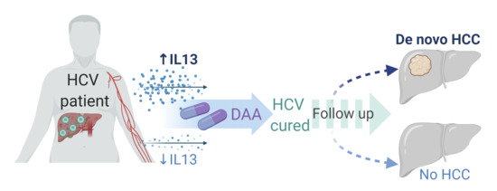
Cancers, Vol. 12, Pages 3820: Circulating IL-13 Is Associated with De Novo Development of HCC in HCV-Infected Patients Responding to Direct-Acting Antivirals

Alexandros G.Sfakianakis shared this article with you from Inoreader
Μέσω Cancers

cancers-12-03820-ag-550.jpg

Cancers, Vol. 12, Pages 3820: Circulating IL-13 Is Associated with De Novo Development of HCC in HCV-Infected Patients Responding to Direct-Acting Antivirals

Cancers doi: 10.3390/cancers12123820

Authors: Zuzana Macek Jílková Arnaud Seigneurin Celine Coppard Laurissa Ouaguia Caroline Aspord Patrice N. Marche Vincent Leroy Thomas Decaens

Direct-acting antivirals (DAAs) are highly effective in targeting hepatitis C virus (HCV) infections, but the incidence of HCV-related hepatocellular carcinoma (HCC) remains still high. In this study, we investigated a cohort of HCV-infected patients treated with DAAs who were followed up for 4 years after sustained virological response (SVR) achievement. Patients who developed de novo HCC following DAA treatment were compared to matched controls who did not develop HCC. These control patients were selected based on DAA treatment, sex, age, fibrosis status, and platelet counts. We evaluated serum levels of 30 immune mediators before, during, at the end of, and three months after DAA treatment using Luminex technology. We identified the immune factors associated with de novo HCC occurrence following DAA treatment. Specifically, interleukin (IL)-4 and IL-13 levels were significantly higher before start of the DAA treatment in the serum of patients who later developed HCC than in co ntrols and stayed higher at each subsequent time point. Least absolute shrinkage and selection operator (LASSO) regression revealed IL-13 as the only strong factor associated with HCC development in this cohort of HCV patients. The difference was observed already at baseline of DAA treatment, which confirms the existence of a specific immune profile in these patients who later develop HCC.

View on the web

Antibiotics, Vol. 9, Pages 920: A Risk Prediction Flowchart of Vancomycin-Induced Acute Kidney Injury to Use When Starting Vancomycin Administration: A Multicenter Retrospective Study

Alexandros G.Sfakianakis shared this article with you from Inoreader
Μέσω Antibiotics

antibiotics-09-00920-g001-550.jpg

Antibiotics, Vol. 9, Pages 920: A Risk Prediction Flowchart of Vancomycin-Induced Acute Kidney Injury to Use When Starting Vancomycin Administration: A Multicenter Retrospective Study

Antibiotics doi: 10.3390/antibiotics9120920

Authors: Takayuki Miyai Shungo Imai Hitoshi Kashiwagi Yuki Sato Shota Kadomura Kenji Yoshida Eri Yoshimura Toshiaki Teraya Takashi Tsujimoto Yukari Kawamoto Tatsuya Itoh Hidefumi Ueno Yoshikazu Goto Yoh Takekuma Mitsuru Sugawara

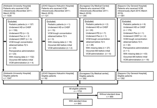
We previously constructed a risk prediction model of vancomycin (VCM)-associated nephrotoxicity for use when performing initial therapeutic drug monitoring (TDM), using decision tree analysis. However, we could not build a model to be used at the time of initial administration due to insufficient sample size. Therefore, we performed a multicenter study at four hospitals in Japan. We investigated patients who received VCM intravenously at a standard dose from the first day until the initial TDM from November 2011 to March 2019. Acute kidney injury (AKI) was defined according to the criteria established by the “Kidney disease: Improving global outcomes” group. We extracted potential risk factors that could be evaluated on the day of initial administration and constructed a flowchart using a chi-squared automatic interaction detection algorithm. Among 843 patients, 115 (13.6%) developed AKI. The flowchart comprised three splitting variables (concomitant drugs (va sopressor drugs and tazobactam/piperacillin) and body mass index ≥ 30) and four subgroups. The incidence rates of AKI ranged from 9.34 to 36.8%, and they were classified as low-, intermediate-, and high-risk groups. The accuracy of flowchart was judged appropriate (86.4%). We successfully constructed a simple flowchart predicting VCM-induced AKI to be used when starting VCM administration.

View on the web

Exceptional Regression of Malignant Pleural Mesothelioma with Pembrolizumab Monotherapy

Alexandros G.Sfakianakis shared this article with you from Inoreader

1260338

The lead author with clinical stage I malignant pleural mesothelioma, epithelioid type, highly programmed cell death ligand 1 (PD-L1) positive, and BAP1 negative, experienced a prompt and exceptionally favorable response to pembrolizumab monotherapy. After cessation of treatment due to immune-related endocrinopathies, complete metabolic response on interim PET/CT scan was achieved. Two years after initial diagnosis, un ifocal tumor reactivation was addressed with successful pembrolizumab monotherapy rechallenge. Immunotherapy, typically not used as frontline treatment for malignant pleural mesothelioma, may provide an effective and durable response for some patients. Based on this single case study, epithelioid type tumors with strongly positive PD-L1 and BAP1-negative immunohistochemical markers may be well suited for treatment with immune checkpoint inhibitors such as pembrolizumab.
Case Rep Oncol 2020;13:1483–1489
View on the web

Pembrolizumab-Induced Severe Neuropathy in a Patient with Metastatic Urothelial Carcinoma after Achieving Complete Response: Guillain-Barré Syndrome-Like Onset

Alexandros G.Sfakianakis shared this article with you from Inoreader

1262137

An 85-year-old female was admitted to our hospital for left ureteral cancer and para-aortic lymph node metastasis. To control hematuria, a laparoscopic retroperitoneal nephroureterectomy was performed, and papillary urothelial carcinoma (pT3b) was found. To treat para-aortic lymph node metastasis, she received chemotherapy with gemcitabine and nedaplatin. After 2 cycles, a computed tomography scan revealed its disappe arance; however, bilateral lung metastases appeared. The patient was administered second-line therapy with pembrolizumab every 3 weeks. After 3 courses, lung metastases disappeared and she achieved a complete response. After the fifth administration of pembrolizumab, she was readmitted with right upper limb pain and weakness in both lower extremities. She was diagnosed with pembrolizumab-induced grade 3 peripheral neuropathy with Guillain-Barré syndrome-like onset. High-dose monocorticotherapy was initiated for treatment. Three weeks later, the pain and weakness of the limbs improved. After discharge, the dose of prednisolone was tapered and there was no relapse of adverse events. Pembrolizumab was discontinued at the onset of neuropathy, but she maintained a complete response.
Case Rep Oncol 2020;13:1490–1494
View on the web

Cranial Nerve Metastasis Treated with Radiotherapy and Chemotherapy in Patients with Lung Adenocarcinoma

Alexandros G.Sfakianakis shared this article with you from Inoreader

1261891

The incidence of central nervous system metastasis is known to be high among patients with lung cancer. The frequency of brain metastasis and carcinomatous meningitis during the entire clinical course of non-small cell lung cancer is reported to be about 40% and 5%, respectively. In contrast, the incidence of cranial nerve metastasis is extremely rare, and detailed reports of its clinical course remain limited. Herein , we report 2 patients diagnosed with cranial nerve metastasis of lung adenocarcinoma and treated with radiotherapy and systemic chemotherapy. Both patients had cranial nerve symptoms, and brain magnetic resonance imaging showed cranial nerve enhancement. However, no evidence of carcinomatous meningitis was noted on magnetic resonance imaging and cerebrospinal fluid cytology. Based on these observations, these patients were diagnosed with cranial nerve metastasis of lung adenocarcinoma. Radiotherapy and chemotherapy were performed in both cases. In both cases, neurological symptoms had not worsened and imaging findings did not indicate any deteriorations. Therefore, radiotherapy and systemic chemotherapy should be considered when treating cranial nerve metastasis of lung adenocarcinoma. Early therapeutic intervention may lead to attenuation of the cranial nerve dysfunction resulting from cranial nerve metastasis.
Case Rep Oncol 2020;13:1495–1500
View on the web

Bilateral Encrusted Metallic Stent Successfully Removed by Ureteroscopic Lithotripsy Using a Ho:YAG Laser in a Patient with Malignant Myeloma

Alexandros G.Sfakianakis shared this article with you from Inoreader

1262257

Ureteral stent encrustation is sometimes encountered, especially in cases in which a ureteral stent has been forgotten. An 84-year-old female patient with malignant myeloma underwent metallic ureteral stent insertion to treat malignant ureteral obstruction. At the time of scheduled ureteral stent exchange, the stent was heavily encrusted and could not be removed on either side. We performed endoscopic lithotripsy to r emove the encrusted ureteral stents. The bilaterally encrusted metallic ureteral stents were successfully removed using Ho:YAG laser lithotripsy after inserting another ureteral stent placement besides the encrusted metallic ureteral stents.
Case Rep Oncol 2020;13:1501–1505
View on the web

Successful Treatment of Advanced Thymic Carcinoma with Carboplatin plus nab-Paclitaxel and Maintenance Monotherapy with nab-Paclitaxel: Two Case Reports

Alexandros G.Sfakianakis shared this article with you from Inoreader

1262234

A standard chemotherapy regimen for advanced thymic carcinoma has not yet been established. We treated 2 cases of thymic carcinoma with carboplatin plus nanoparticle albumin-bound (nab)-paclitaxel, and nab-paclitaxel maintenance therapy. The first case was a 68-year-old female, admitted for dyspnea and left shoulder pain. Chest computed tomography (CT) showed a huge mass in the anterior mediastinum, pleu ral and pericardial effusions, and multiple lung metastases. Specimens obtained from the anterior mediastinal mass by CT-guided needle biopsy revealed squamous cell carcinoma of the thymus, which was in stage IVB. The patient was administered carboplatin plus nab-paclitaxel as first-line treatment. After 3 cycles of chemotherapy, a partial response was observed with marked shrinkage of the tumor. Following 6 cycles of chemotherapy, nab-paclitaxel maintenance therapy was initiated. Disease progression was seen 9.1 months after initiation of treatment. The patient experienced no serious adverse events. The second case was a 70-year-old male who had productive cough, dyspnea, and right-sided chest pain. Chest CT revealed a huge mass in the anterior mediastinum, pericardial effusion, and multiple lymphadenopathies. Specimens obtained from station 11s by endobronchial ultrasound-guided transbronchial needle aspiration revealed undifferentiated thymic carcinoma, which was in stage IVB. Six cycles of carboplatin plus nab-paclitaxel were administered, followed by 5 cycles of nab-paclitaxel for maintenance. A partial response was seen, which was sustained for more than 13 months. The patient experienced no serious adverse events. These cases show that chemotherapy with carboplatin plus nab-paclitaxel and nab-paclitaxel as maintenance therapy can be a useful regimen for advanced thymic carcinoma.
Case Rep Oncol 2020;13:1506–1512
View on the web

Hepatic Inflammatory Myofibroblastic Tumor Detected in the Fetal Period That Caused an Oncologic Emergency

Alexandros G.Sfakianakis shared this article with you from Inoreader

1262247

A huge abdominal cystic lesion with ascites was detected in a male neonate at 31 weeks of gestation. Increasing ascites and the appearance of subcutaneous edema were detected, which caused fetal hydrops. The patient was delivered by emergency cesarean section at 33 weeks of gestation. The birth weight was 2,407 g, and the Apgar score was 8/9 points (1-/5-min values). Breathing at birth was stable, but the patient pres ented with remarkable abdominal distention due to the ascites. Later, the patient presented with tachypnea, and breathing gradually worsened, so an emergency operation was performed. There were no intraoperative findings within the small intestine, but there was a large amount of ascites and a cystic mass arising from the liver. The patient's breathing and circulation dynamics could only be stabilized by ascites removal, so only a tumor biopsy was performed. The pathological findings led to the diagnosis of an inflammatory myofibroblastic tumor, and steroids were administered early after surgery for the purpose of an anti-inflammatory effect and tumor shrinkage. The abdominal distention was alleviated, and blood examinations showed a reduced inflammatory response. There was no apparent shrinkage of the tumor, however; thus, radical surgical treatment was performed on day 24. The postoperative course was uneventful, so the patient was discharged on day 36. Seven years after the ope ration there has been no recurrence or distant metastasis.
Case Rep Oncol 2020;13:1513–1519
View on the web

High-Dose Omalizumab versus Ligelizumab for the Treatment of Chronic Spontaneous Urticaria: Do Not We Need a Head-To-Head Comparison?

Alexandros G.Sfakianakis shared this article with you from Inoreader

?image=000512373-1.jpg


Int Arch Allergy Immunol
View on the web